Digital radiography (DR) is an advanced form of x-ray inspection which produces a digital radiographic image instantly on a computer. This technique uses x-ray sensitive plates to capture data during object examination, which is immediately transferred to a computer without the use of an intermediate cassette. The incident x-ray radiation is converted into an equivalent electric charge and then to a digital image through a detector sensor.
Compared to other imaging devices, flat panel detectors, also known as digital detector arrays (DDAs) provide high quality digital images. They can have better signal-to-noise ratio and improved dynamic range, which, in turn, provides high sensitivity for radiographic applications.
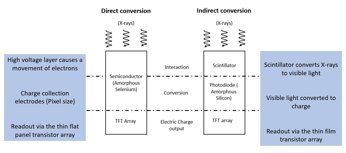
Flat panel detectors work on two different approaches, namely, indirect conversion and direct conversion. Indirect conversion flat panel detectors have a scintillator layer which converts x-ray photons to photons of visible light and utilise a photo diode matrix of amorphous silicon to subsequently convert the light photons into an electrical charge. This charge is proportional to the number and energy of x-ray photons interacting with the detector pixel and therefore the amount and density of material that has absorbed the x-rays.
Direct conversion flat panel detectors use a photo conductor like amorphous selenium (a-Se) or Cadmium telluride (Cd-Te) on a multi-micro electrode plate, providing the greatest sharpness and resolution. The information on both types of detectors is read by thin film transistors.
In the direct conversion process, when x-ray photons impact over the photo conductor, like amorphous Selenium, they are directly converted to electronic signals which are amplified and digitised. As there is no scintillator, lateral spread of light photons is absent here, ensuring a sharper image. This differentiates it from indirect construction.
When a flat panel detector is coupled with an appropriate manipulator and image processing software, they can be used for x-ray Computed Tomography (CT) inspection, producing a 3D image of the external and internal structure of the test object.
Another type of digital x-ray detection media are linear detector arrays (LDAs). These consist of a single row of x-ray detection pixels, rather than a matrix. The LDA or the object under inspection need to move relative to each other to formulate a 2D radiographic image. LDAs are suited to inspection of objects moving on a conveyor belt.
Find out TWI's capabilities
Digital radiography offer many advantages to the non-destructive testing community, including:
- Shorter exposure times
- Real-time applications
- Use of analysis tool and defect recognition software
- Improved detail detectability
- Enhanced SNR and linearity
- Reduced inspection time, as no chemical processing of film is required
- Eliminates processing chemical, hence safe for environment
- Digital image enhancement and data storage
- Higher productivity
- Portability
- Increased dynamic range enables multiple thickness to be inspected in one shot
- Immediate feedback
- Easy to transfer to customers electronically
Digital radiography systems use active matrix flat panels or linear detector arrays, which consist of a detection layer deposited over an active matrix array of thin film transistors and photodiodes. Digital radiography images are converted to digital data in real-time and are available for analysis within seconds.
Instead of traditional x-ray film, computed radiography cassettes use photo-stimulated luminescence screens to capture the X-ray image. The computed radiography cassette goes into a reader, which converts the stored data into a digital image. Computed radiography imaging plates are flexible and do not require a rigid holder. Flexible cassettes are available that enable the detector to be fitted into curved areas.
While both computed radiography and digital radiography have a wider dose range and can be post processed to eliminate noise, DR has many advantages over computed radiography. Digital radiography improves workflow by producing higher image quality instantaneously while providing up to three times more dose efficiency than computed radiography. With ongoing technological advancements and reduction in price, digital radiography is fast becoming the preferred choice for non-destructive testing operators.
As with all NDT processes, digital radiography as numerous applications, including:
- Aerospace product examination
- Detection of Corrosion Under Insulation (CUI) in petrochemical, oil and gas and power generation industries
- Detection of Flow accelerated corrosion
- Foreign object detection
- Casting and weld inspection
- Inspection of composites and fibre reinforced components
- Product and process development
How can TWI Help?
TWI has a long history of working with its Members, across a range of industry sectors, on non-destructive testing processes such as digital radiography. TWI offers digital radiographic inspection services as well as bespoke digital radiography system prototyping and consultancy.
Please contact us to learn more.
contactus@twi.co.uk